Dental Issues in Reptiles
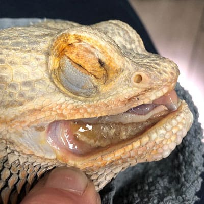
Lizards have lips, gums, and salivary glands, just like mammals do. But they also have mucous glands which, together with the salivary glands, help them lubricate and swallow their food. In older reptiles like bearded dragons and chameleons, the teeth are more delicate and prone to being worn down and damaged. Turtles and snakes are more susceptible to infections and inflammation of the mouth, and stomatitis, which causes painful sores and swelling in the oral cavity.
These dental problems are typically caused by poor husbandry, especially a diet that is lacking in calcium and vitamin A. Without these crucial vitamins, your reptile is more prone to secondary oral infection. Live prey feedings can also open the door to bacteria and fungus.
What Causes Dental Disease?
Exotic pets can develop dental disease as the result of a congenital trait, or they can acquire it during the course of their life.
Acquired causes of dental disease generally include:
- Improper nutrition
- Insufficient UVB radiation
- An enclosure that is too small and/or contains the wrong materials
- Parasites like nematodes and flukes (reptiles)
- Trauma
- Systemic disease
- Neoplasia (cancer)
Clinical Signs of Dental Disease in Exotics
Exotic pets generally will hide any clinical signs of illness or pain until they are noticeably debilitated. Therefore, a pet’s clinical signs tie in directly to the severity of their condition. Mild disease will likely not yield any signs. But once a tooth is affected, with time the surrounding teeth will also be affected.
Clinical Signs We Look For Include:
- Difficulty eating and/or decreased appetite
- Anorexia
- Changes in fecal output, size, and appearance
- Nasal discharge
- Increased tearing or eye bulging
- Facial masses/swellings (could be a sign of an abscess)
- Bruxism (teeth grinding, often a sign of pain)
Clinical Signs In Reptiles:
- Drooling
- Decreased appetite
- Inability to hold the mouth closed properly
- Change in lip margin
- Lip deformities
- Oral discharge
- Jaw swelling
How We Diagnose Dental Disease
We take several crucial steps to make the most accurate diagnosis possible and properly stage the disease process:
- Physical examination
- Endoscopic oral examination with pet under sedation
- Blood analysis (recommended)
- Culture and sensitivity of affected areas
- Dental radiography (essential for any pet with suspected dental disease; allows us to view the teeth in their entirety, not just the 20-30% visible above the gumline)
- Skull/dental CT scans (more sensitive than radiographs and allow for better evaluation of the nasal passages, spaces behind the eyes, and middle ear structures)
Treating and Preventing Dental Disease
Depending on the nature of your pet’s dental disease, we can take several different approaches to treating and managing their condition so they can live a better life.
Teeth Trimming
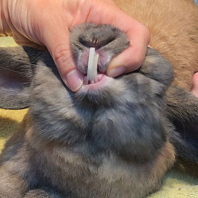
Crown height reduction (teeth trimming) and diet correction in the early stages of the disease can help to reverse the damage in rabbits and pocket pets. For pets with a moderate or severe case of disease, teeth trimming will need to be done repeatedly. We use a dental bur and trimming forceps to trim overgrown incisors and cheek teeth. No other tools should be used for this procedure, as they can damage the teeth and cause abnormal regrowth and/or infection.
Husbandry Optimization
For dental disease cases ranging from mild to severe, reptiles and other exotics need optimal living conditions to heal as fully as possible from their condition. A healthy, appropriate diet, correct temperature and humidity, and quality enclosure materials are all essential.
For rabbits we recommend unlimited, high-quality grass hay such as timothy, brome, or orchard, and various fibrous, leafy green vegetables. Adult animals do not need pellets, but if you choose to give pellets to your pet, make sure they are timothy hay-based instead of alfalfa-based, with no additives. Do not give your pet more than 1-2 tbsp of pellets daily.

Antimicrobial Therapy and Scaling
Under sedation, your pet’s oral cavity will need to be thoroughly flushed with an antimicrobial solution. If there is any necrotic or devitalized tissue present, it will need to be debrided. Any calculus present will be removed with dental scaling equipment. For severe dental disease cases, the pet will need to be tube fed to bypass their oral cavity entirely. Topical antimicrobial therapy is also required throughout treatment, along with systemic antibiotics and pain relief medication.
Tooth Extraction
Another treatment option is to extract an affected tooth with the patient under general anesthesia. Teeth that are loose, infected/abscessed, fractured, or severely maloccluded should be extracted to prevent further damage to your pet’s mouth. We can perform extractions intra-orally or extra-orally, depending on the size of the pet and how we can access the tooth.
Our goal is to remove the cause of the infection, which tends to be an infected tooth (or teeth), or infected bone. After the extraction, we may need to employ further, repeated treatments like lancing and flushing, systemic antibiotics, complete surgical excision, and antibiotic bead impregnation to fully treat the infection. These procedures are painful, so we will provide your pet with complete pain management and nutritional support before, during, and after.


